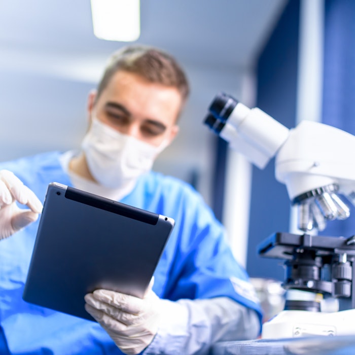
Scientists developed a smartphone microscope to help skin professionals spot skin cancer, especially in remote locations. The effort is to detect at least some of the two and three million non-melanoma skin cancers and 132,000 melanoma skin cancers each year.
According to the report in the Archives of Pathology & Laboratory Medicine, a smartphone microscope can be made with a three mm ball lens, which is a piece of plastic normally used for laser optics. The lens is placed over the smartphone lens with tape to hold everything in place.
Proper Use
To utilize the smartphone microscope, a skin sample is placed on a slide, and the smartphone microscope is held over the sample until the image comes into focus. Once the sample is in focus, an image can be taken to send to a dermatologist.
“Doctors in some remote areas don’t have access to the high-powered microscopes we use to evaluate skin samples,” said Richard Jahan-Tigh, study lead author and assistant professor at The University of Texas Health Science Centre at Houston, US. “Doctors there could conceivably use their smartphones to photograph growths and forward them for examination.”
The Smartphone Microscope Results
The scientists examined 1,021 slides of specimens for the study. According to Jahan-Tigh, the smartphone microscope resulted in detecting about 90% of the non-melanoma skin cancers and 60% for the melanoma skin cancers.
“This is a good first step to show that smartphone microscopy has a future in dermatology and pathology,” said Jahan-Tigh.
Source: The Health Site