Melasma is a subset of a general condition in the skin called hyperpigmentation. It is important to understand how skin develops hyperpigmentation, as well as to become familiar with the general types in order to manage the condition with the proper modalities. This article deals with the various root causes of nondisease-oriented hyperpigmentation and treatment methods to utilize, as well as treatments to avoid, depending on the specific type.
Skin color is developed by melanocytes: color-producing cells in the skin that determine different types and colors of melanin. Melanosomes are color organelles, or subunits, that are transferred from the melanocytes to the keratinocytes, which live in epidermal skin cells. The color of a person’s skin is largely dependent on a variety of factors including, but not exclusive to:
- The amount of melanin produced;
- The ratios of the type of melanin;
- The size of the melanosome; and
- The distribution of melanin throughout skin.
Pigmentation disorders
Aesthetically pleasing skin—no matter what color—exhibits an even distribution of melanin throughout. Skin that has even tonality is pleasing to the eye and contributes to the common idea of what is perceived to be beautiful. There are several pigmentation disorders that arise in skin, leading to uneven tone.
Oxidative damage. Oxidative damage commonly results due to UV light exposure from the sun, and takes the form of discrete spots on skin, which are often concentrated on the forehead, and across the cheeks and nose. Ingredients, such as endonucleases, can aid in improving this kind of hyperpigmentation.
Post-inflammatory hyperpigmentation (PIH). PIH often results from active cystic acne and after laser or intense pulsed light (IPL) treatments, as well as other mechanically induced skin trauma. The inflammation produced from these and other sources leads to higher localized melanin production in the skin, which results in hyperpigmentation.
Native freckles and ethnic melanin banding. Native freckles and ethnic melanin banding are genetically linked root causes of hyperpigmentation. Although it is sometimes difficult to distinguish between the two, a trained skin care professional can do this. Melanin banding in skin usually presents itself as smooth, darker areas around the eyes and chin.
Melasma. Melasma, also known as chloasma or pregnancy mask, is a hormone-induced condition. The onset is often consistent with hormone spikes that occur during pregnancy; changes in birth control; shifts in estrogen, estradiol and progesterone; and, in lesser cases, high stress-related life issues. The presence of melasma is marked by symmetrical map-like lesions on the face, typically in the cheek and forehead areas, but often appearing on the upper lip.
There are two primary types of melasma: epidermal melasma and dermal melasma, and they are distinctly different in diagnosis and treatment. Epidermal melasma can be observed in the epidermal tissue and is generally tan-to-brown in color. Dermal melasma is in the dermal tissue and appears gray-to-grayish brown in color. A definitive method to determine the difference between epidermal and dermal melasma is to take photos of the skin with color UV reflectance photography.
Case Study 1. See Case Study 1: UV Imaging. Notice how apparent the melasma becomes when overlaying the two images. It can be seen that this client is exhibiting primarily epidermal melasma. The melasma will appear in the standard and the color UV imaging if it is epidermal, but will not if it is dermal melasma.
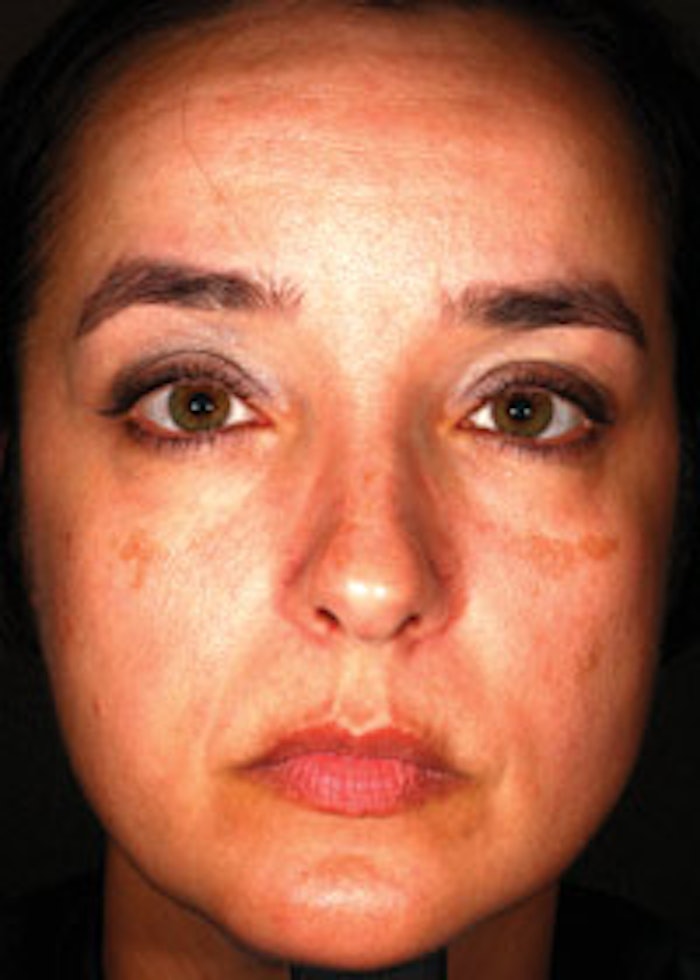
Case Study 2. You can see in Image 1 a client presented with two symmetrical hyperpigmented lesions. At first glance, the case could be made for melasma since it presents as a map-like symmetrical pattern, but this is PIH. The history of the client indicated that her sun damage was over-treated with IPL, which caused a significant inflammation response. There were several more interventions with IPL, continually worsening the condition. Upon client referral, image analysis, biometric and colorimetric measurements were taken. It was clear though underlying erythema (redness) measurements, as well as color UV imaging, that PIH was the root cause of this hyperpigmentation. Once this was determinationed, a few decisions were immediately made.
- No thermal or chemical treatments, such as laser, IPL or peels, should be performed. These modalities would only further inflame the area and worsen the PIH.
- Exposure to UV light and environmental oxidative factors, such as cigarette smoke, should be strictly avoided and a broad-spectrum SPF 50 should be used daily.
- A topical regimen, including skin-lightening agents and anti-inflammatories, as well as barrier repair agents, is the preferred course of treatment.
In Image 2, you can see the result of a course of this type of topical treatment. The treatment included a glycolic-based cleanser with pH 3.5–4.0 to encourage desquamation of the corneocytes; an anti-inflammatory serum with barrier repair actives formulated to reduce multiple pathways of inflammation; a formulated lightening product including active ingredients for PIH suppression; as well as 4% hydroquinone, and a moisturizing treatment lotion for hydration purposes. This course of treatment took six months.
Case Study 3. The second client can be seen in Case Study 3: Epidermal Melasma. This client presented with dark map-like areas on the cheeks, forehead and upper lip. The images show the hyperpigmented areas in color UV reflected light imaging, which confirms epidermal melasma with little dermal melasma present. This is a confirmed case of epidermal melasma. Client history indicates a 10-year progressive history of active melasma with birth control use for more than 25 years.
The approach taken with this client was the use of topically formulated products, along with a series of medical-grade peels. It is important to understand the condition of the skin before applying topical ingredients and peels. This client did not have an impaired barrier, and there was little inflammation present; therefore, an aggressive peel regimen was prescribed. The peel included 2% hydroquinone, as well as lactic, kojic, salicylic and citric acids. A series of three peels, four weeks apart was undertaken. The topical regimen included a glycolic acid cleanser to maintain a regular desquamation pattern; a hydrating 4% hydroquinone lotion formulated specifically with a melanocyte-stimulating hormone inhibitor; a tyrosinase inhibitor; and a melansome transfer inhibitor. The results of this treatment can be seen in Case Study 3: Aggressive Peels.
Many variables
There are many variables when dealing with hyperpigmentation—in particular, melasma. The proper diagnosis is critical in clearing the client and not worsening the condition. As was evidenced in Case Study 1, if chemical or light therapy was used, the probability of worsening the client’s condition was high. It is also critical that the topical regimen is appropriate for the client.
REFERENCE
(Accessed Dec 10, 2013)

Editor’s note: Don’t miss Robert Manzo discussing this topic live during the Advanced Education Conference Program at Face & Body® Midwest in Chicago on Saturday, March 22, 2014. Register today at www.FaceandBody.com/midwest!